You are not logged in. (Sign In)
Northwestern University Schizophrenia Data and Software Tool (NUSDAST)
PI: Wang
Summary:
To make structural magnetic resonance (MR) imaging data, genotyping data, and neurocognitive data as well as analysis tools available to the schizophrenic research community.
Please note that this dataset is also accessible through the SchizConnect project at: http://schizconnect.org. Here, you will be able to get additional schizophrenia neuroimaging data from FBIRN and COINS.
External Link: Northwestern University Schizophrenia Data and Software Tool (NUSDAST)
Papers:
- Heritability of Multivariate Gray Matter Measures in Schizophrenia .
Turner, J. A., Calhoun, V. D., Michael, A., van Erp, T. G. M., Ehrlich, S., Segall, J. M., Gollub, R. L., Csernansky, J., Potkin, S. G., Ho, B.-C., Bustillo, J., Schulz, S. C., Fbirn, n., Wang, L.
Twin Research and Human Genetics 2012; 15: 324-335
View Article
- Cortical thickness in neuropsychologically near-normal schizophrenia.
Cobia, D. J., Csernansky, J. G., and Wang, L.
Schizophr Res 2011; 133: 68-76
View Article
- Northwestern University Schizophrenia Data and Software Tool (NUSDAST).
Wang, Lei, Alexander Kogan, Derin Cobia, Kathryn Alpert, Anthony Kolasny, Michael I. Miller, Daniel Marcus
Frontiers in Neuroinformatics 2013; 7:25
View Article
- SchizConnect: Mediating neuroimaging databases on schizophrenia and related disorders for large-scale integration.
Wang, L., Alpert, K.I., Calhoun, V.D., Cobia, D.J., Keator, D.B., King, M.D., Kogan, A., Landis, D., Tallis, M., Turner, M.D., Potkin, S.G., Turner, J.A., Ambite, J.L.
NeuroImage 2016; 124: 1155-1167
View Article
- Northwestern University schizophrenia data sharing for SchizConnect: A longitudinal dataset for large-scale integration.
Kogan A, Alpert K, Ambite JL, Marcus DS, Wang L
NeuroImage 2016; 124: 1196-1201
View Article
Presentations:
- SchizConnect: Large-Scale Schizophrenia Neuroimaging Data Integration and Sharing
Lei Wang, Jose Luis Ambite, Jessica Turner, Steven Potkin NIMH (NIMH, Bethesda, MA -- Sep 2014)
Download Presentation
- Large-Scale Schizophrenia Neuroimaging Data Integration and Sharing
Wang L, Alpert KI, Calhoun V, Keator D, King M, Kogan A, Landis D, Tallis M, Potkin SG, Turner JA, Ambite JL Annual Meeting of the American College of Neuropsychopharmacology (ACNP) (Phoenix, Arizona -- December 2011)
Download Presentation
Funding:
- 1 R01 MH084803 NIMH: Schizophrenia Data and Software Tool Federation using BIRN Infrastructure (PI: Wang) - To make structural magnetic resonance (MR) imaging data, genotyping data, and neurocognitive data as well as analysis tools available to the schizophrenic research community. (07/01/2009 - 12/31/2012).
Details:
Access
To determine if our data can help your research, please visit SchizConnect and see what we have available. Guests can specify search parameters to isolate the data that meets their needs.
Data dictionary
A data dictionary can be obtained here.
To access NUSDAST, the following are required:
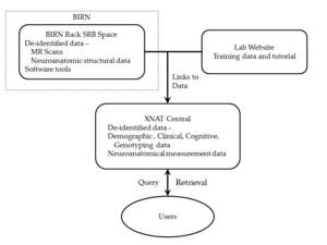
1. Obtain an account on XNAT Central;
2. Request access to NU Schizophrenia Data and Software Tool Federation using BIRN Infrastructure (NUSDAST) from XNAT Central account;
3. Sign the NUSDAST Data Use Agreement.
Background
In collaboration with John G. Csernansky, MD, Lizzie Gilman Professor and Chair of Psychiatry and Behavioral Sciences at Northwestern Feinberg School of Medicine, Michael I. Miller, PhD, Professor of Biomedical Engineering and Director of the Center for Imaging Science at the Johns Hopkins University, and Daniel Marcus, PhD, Research Assistant Professor of Radiology at Washington University School of Medicine, and Director of the Neuroinformatics Research Group at Washington University, we are making structural magnetic resonance (MR) imaging data, genotyping data, and neurocognitive data as well as analysis tools available to the schizophrenic research community.
Sharing data and tools across a research community adds tremendous value to the efforts of that community. The project, entitled “Schizophrenia Data and Software Tool Federation using Biomedical Informatics Research Network (BIRN) Infrastructure,” is funded by the National Institute of Mental Health, using the BIRN as a platform for data sharing and informatics tool sharing that extends beyond the neuroimaging researchers involved in the test beds, to include other areas of neuroscience beyond imaging, and to include biomedical research beyond neuroscience.
As part of the growing field of Computational Anatomy (CA), our group has developed tools to identify and characterize brain structural abnormalities in schizophrenia, some of which meet the criteria for disease endophenotypes. In this effort, we have collected high resolution magnetic resonance (MR) datasets from more than 270 subjects using the same MR scanner platform and sequences. Longitudinal MR data are also available on a majority of these subjects. Using CA tools, we have generated surface maps for all of the deep subcortical structures (i.e., hippocampus, amygdala, thalamus, caudate nucleus, nucleus accumbens, putamen and globus pallidus). In addition, smaller datasets of variables related to the volume, thickness and surface area of cortical structures (e.g., the cingulate gyrus) have been generated. Finally, we have constructed manual segmentation datasets for all these structures, which can be used for the validation of new computational methods.
When made publically accessible, these high resolution scans and the associated structural data will be invaluable to the neuroscience community in many ways. First, other groups of scientists will be able to use these data to generate or test new hypotheses related to the maldevelopment of brain structures and neural networks in individuals with schizophrenia. Second, scientists in other groups would be able to rapidly replicate findings produced using their own datasets. Third, the data could be used to test and validate new brain mapping tools. Further, the CA pipeline designed for the analysis of these datasets, which consists of landmarking and diffeomorphic mapping tools along with training and validation datasets, will enable others to study other MR datasets collected from other clinical samples. A key group of intended users of our distribution is researchers who may have their own imaging data but lack software tools for mapping subcortical brain structures. In that group, our software tools would be needed and used on our training data, therefore enabling these researchers to obtain structural measurements in their images.
